Blood Vessels Labeled Brain - Arterial Supply Of The Brain Circle Of Willis Geeky Medics : Equal to the intestinal muscles that move the food morsel along brain level:
Blood Vessels Labeled Brain - Arterial Supply Of The Brain Circle Of Willis Geeky Medics : Equal to the intestinal muscles that move the food morsel along brain level:. The dense tight junctions between endothelial cells prevent paracellular transport through the. The blood vessels are the components of the circulatory system that transport blood throughout the human body. Blood in the brain is supplied by two pairs of large blood vessels (arteries): If you use this material, please attach a link to the artwork, i would really love to see it :d. The 500 ms patients, both adults and children, also underwent mri scans of the brain to measure iron deposits in surrounding areas of the brain.
Equal to the intestinal muscles that move the food morsel along brain level: This is particularly important structure due to its clinical implications, which are discussed in more detail in the article. The brain and its surrounding blood vessels exist in a close relationship. Supplies the posterior brain, blood supply to the entire brain is ensured by anastomoses between the vessels. Blood vessels in red in close communication with proliferating neuronal cells in the mouse cortex at embryonic day 10.
Duke Neurosciences Lab 1 Surface Anatomy Of The Brain from brain.oit.duke.edu The blood vessel wall is endowed with connective tissue, smooth muscle, and striated muscles. Researchers have discovered how cells of the blood vessels sense the metabolic condition of the brain and alter vascular function in response. In the cerebral medulla, the arteries and veins of the right side of the body are controlled from the left side of the brain; However, detecting vessels is still a challenging task those labeled as background or vessel voxels are excluded from consideration in later computation. The brain and its surrounding blood vessels exist in a close relationship. This vessel supplies blood to the front part of your brain, knows as your frontal lobe. Using medaka ( oryzias latipes ) as a model, the current protocol presents a quick and direct technique to label blood vessels in brain and pituitary by. Brain vessel segmentation is a fundamental component of cerebral disease screening systems.
He says the restricted vessels prevent the blood from draining fast enough and injure the brain by causing a build up of iron which leads to ms.
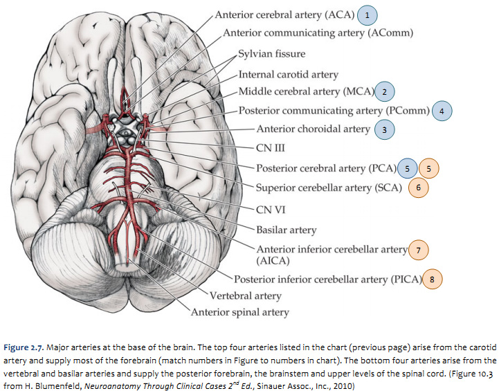
Only some of the vessels that exist in a real brain have been labeled. About 2 years ago updated: This is particularly important structure due to its clinical implications, which are discussed in more detail in the article. Posterior communicating a internal carotid а. Internal carotid artery (anterior circulation), vertebral artery (posterior circulation), and their hexagonal anastomotic network called blood brain barrier refers to the wall between the brain tissue and blood vessels. In the cerebral medulla, the arteries and veins of the right side of the body are controlled from the left side of the brain; The brain and its surrounding blood vessels exist in a close relationship. Towards the anterior side of the brain, those arteries are the internal carotid arteries. In the article on the ventricles within the cns, we will discuss their structure and. The blood vessels are the components of the circulatory system that transport blood throughout the human body. Blood vessels are referred to collectively as the vascular system and, together with the heart, make up the circulatory system or cardiovascular system. There is a right sided aca and a left sided aca. Brain vessel segmentation is a fundamental component of cerebral disease screening systems.
Label the veins of the anterior forearm and hand. This is particularly important structure due to its clinical implications, which are discussed in more detail in the article. The 500 ms patients, both adults and children, also underwent mri scans of the brain to measure iron deposits in surrounding areas of the brain. Towards the anterior side of the brain, those arteries are the internal carotid arteries. The blood vessel wall is endowed with connective tissue, smooth muscle, and striated muscles.
Biology Of The Blood Vessels Heart And Blood Vessel Disorders Msd Manual Consumer Version from www.msdmanuals.com About 2 years ago updated: Comes off the subclavian a., ascends although the internal carotid a. Blood is also supplied to the brain by the vertebral a. This vessel supplies blood to the front part of your brain, knows as your frontal lobe. Microscopically, it is formed by the endothelium of the blood vessel. Using medaka ( oryzias latipes ) as a model, the current protocol presents a quick and direct technique to label blood vessels in brain and pituitary by. Blood vessel endothelium is continuous with the inner tissue lining of organs such as the brain, lungs, skin, and heart. Over 1 year ago version:
Blood vessel endothelium is continuous with the inner tissue lining of organs such as the brain, lungs, skin, and heart.
And these vessels are very small, very fine, and sometimes they clot and produce strokes in the brain stem that can have a variety of consequences. The carotid arteries and the vertebral arteries anterior cerebral artery (aca): Blood vessel endothelium is continuous with the inner tissue lining of organs such as the brain, lungs, skin, and heart. Blood is also supplied to the brain by the vertebral a. • identification of blood vessels as arteries, capillaries or veins from the structure of their walls. Blood in the brain is supplied by two pairs of large blood vessels (arteries): Comes off the subclavian a., ascends although the internal carotid a. The difference in the structural characteristics of arteries, capillaries and veins is attributable to their respective functions. Blood travels from the heart in arteries, which branch into smaller and smaller vessels, eventually becoming arterioles. Fill in the blanks with the appropriate words to describe blood flow from the heart. The brain and its surrounding blood vessels exist in a close relationship. In the article on the ventricles within the cns, we will discuss their structure and. Endothelial cells are labeled in red and pericytes in green.
This vessel supplies blood to the front part of your brain, knows as your frontal lobe. Blood vessels are referred to collectively as the vascular system and, together with the heart, make up the circulatory system or cardiovascular system. All cell types in the blood vessel wall can affect vessel diameter. Brain vessel segmentation is a fundamental component of cerebral disease screening systems. Comes off the subclavian a., ascends although the internal carotid a.
About The Heart And Blood Vessels from api.kramesstaywell.com These vessels transport blood cells, nutrients, and oxygen to the tissues of the body. The 500 ms patients, both adults and children, also underwent mri scans of the brain to measure iron deposits in surrounding areas of the brain. This vessel supplies blood to the front part of your brain, knows as your frontal lobe. Endothelial cells are labeled in red and pericytes in green. Supplies the anterior brain and the vertebral a. Blood vessels in red in close communication with proliferating neuronal cells in the mouse cortex at embryonic day 10. Blood vessel endothelium is continuous with the inner tissue lining of organs such as the brain, lungs, skin, and heart. Microscopically, it is formed by the endothelium of the blood vessel.
The two cell types ensure the integrity of the neural vasculature by maintaining the blood.
The precise relation between blood vessels and brain regions, reflecting the physiology and pathology of brain function directly and accurately, has remained largely unknown. In the cerebral medulla, the arteries and veins of the right side of the body are controlled from the left side of the brain; The two cell types ensure the integrity of the neural vasculature by maintaining the blood. The brain and its surrounding blood vessels exist in a close relationship. Blood vessels are intricate networks of hollow tubes that transport blood throughout the entire body so that it can deliver valuable nutrients to and remove waste from cells. The 500 ms patients, both adults and children, also underwent mri scans of the brain to measure iron deposits in surrounding areas of the brain. They also take waste and carbon dioxide away from the tissues. Equal to the intestinal muscles that move the food morsel along brain level: All cell types in the blood vessel wall can affect vessel diameter. Cerebral arterial circle anterior communicating posterior cerebral a middle cerebral al reset zoom. Supplies the anterior brain and the vertebral a. Using medaka ( oryzias latipes ) as a model, the current protocol presents a quick and direct technique to label blood vessels in brain and pituitary by. Blood is also supplied to the brain by the vertebral a.
The 500 ms patients, both adults and children, also underwent mri scans of the brain to measure iron deposits in surrounding areas of the brain blood vessels labeled. Blood vessels are critical to deliver oxygen and nutrients to all of the tissues and organs throughout the body.


0 Komentar